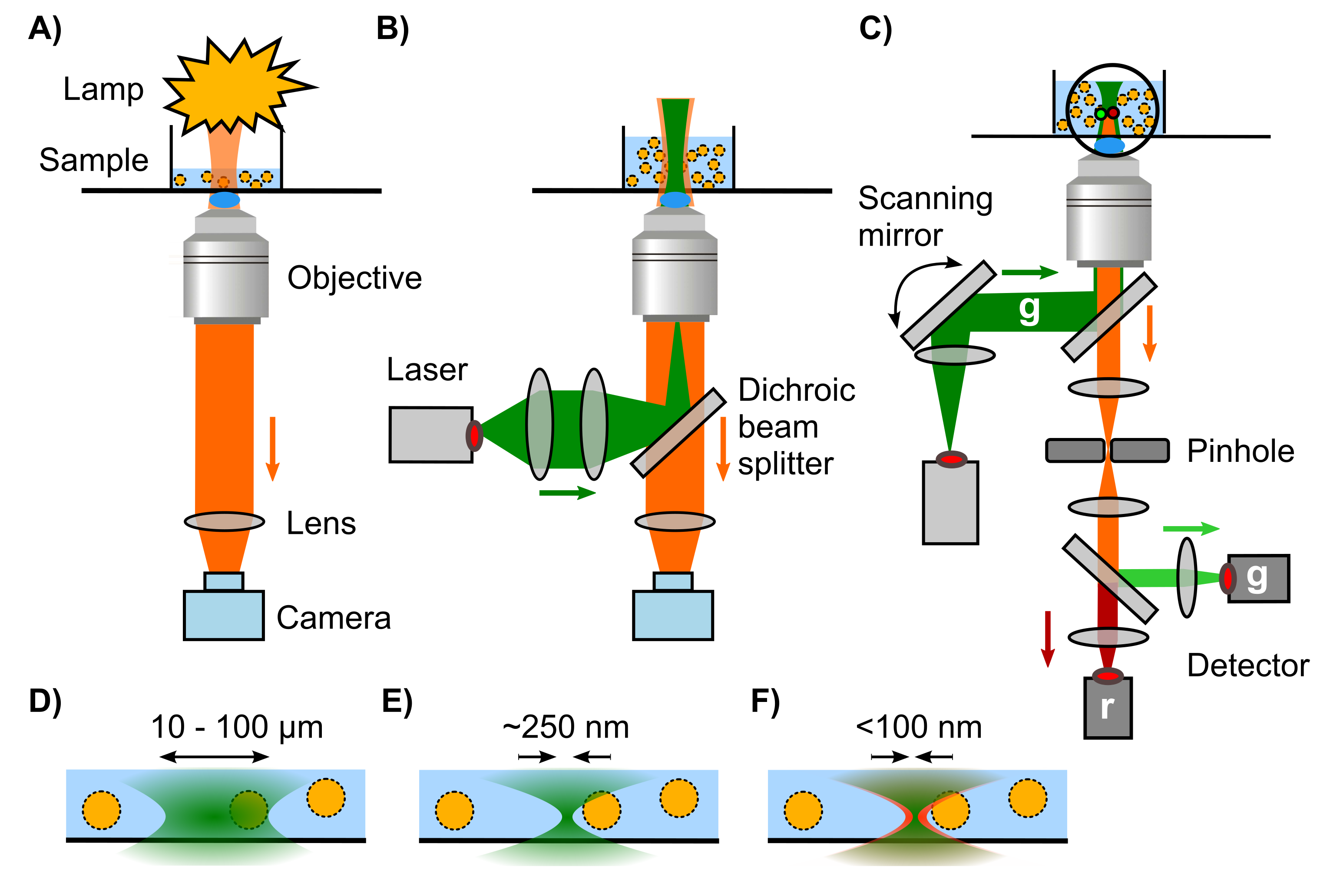
Publication: Visualisation of dCas9 target search in vivo using an open-microscopy framework

K.J.A. Martens, S. van Beljouw, S. van der Els, J.N.A. Vink, S. Baas, G.A. Vogelaar, S.J.J. Brouns, P. van Baarlen, M. Kleerebezem, J. Hohlbein, Nature Communications, 10, 3552, 2019, [link]

CRISPR-Cas9 is widely used in genomic editing, but the kinetics of target search and its relation to the cellular concentration of Cas9 have remained elusive. Effective target search requires constant screening of the protospacer adjacent motif (PAM) and a 30 ms upper limit for screening was recently found. To further quantify the rapid switching between DNA-bound and freely-diffusing states of dCas9, we developed an open-microscopy framework, the miCube, and introduce Monte-Carlo diffusion distribution analysis (MC-DDA). Our analysis reveals that dCas9 is screening PAMs 40% of the time in Gram-positive Lactoccous lactis, averaging 17 ± 4 ms per binding event. Using heterogeneous dCas9 expression, we determine the number of cellular target-containing plasmids and derive the copy number dependent Cas9 cleavage. Furthermore, we show that dCas9 is not irreversibly bound to target sites but can still interfere with plasmid replication. Taken together, our quantitative data facilitates further optimization of the CRISPR-Cas toolbox.

miCubeNatCommun


Pre-print: Direct visualization of native CRISPR target search in live bacteria reveals Cascade DNA surveillance mechanism

J.N.A. Vink, K.J.A. Martens, M. Vlot, R.E. McKenzie, C. Almendros, B. Estrada Bonilla, D.J.W. Brocken, J. Hohlbein, S.J.J. Brouns, bioRxiv, 2019, [link]

CRISPR-Cas systems encode RNA-guided surveillance complexes to find and cleave invading DNA elements. While it is thought that invaders are neutralized minutes after cell entry, the mechanism and kinetics of target search and its impact on CRISPR protection levels have remained unknown. Here we visualized individual Cascade complexes in a native type I CRISPR-Cas system. We uncovered an exponential relationship between Cascade copy number and CRISPR interference levels, pointing to a time-driven arms race between invader replication and target search, in which 20 Cascade complexes provide 50% protection. Driven by PAM-interacting subunit Cas8e, Cascade spends half its search time rapidly probing DNA (∼30 ms) in the nucleoid. We further demonstrate that target DNA transcription and CRISPR arrays affect the integrity of Cascade and impact CRISPR interference. Our work establishes the mechanism of cellular DNA surveillance by Cascade that allows the timely detection of invading DNA in a crowded, DNA-packed environment.

2019_Vink_bioRxiv

Pre-print: An open microscopy framework suited for tracking dCas9 in live bacteria

K.J.A. Martens, S. van Beljouw, S. van der Els, S. Baas, J.N.A. Vink, S.J.J. Brouns, P. van Baarlen, M. Kleerebezem, J. Hohlbein, bioRxiv, 2018 [link]

Super-resolution microscopy is frequently employedin the life sciences, but the number of freely accessible and affordable microscopy frameworks, especially for single particle tracking photo-activation localization microscopy (sptPALM), remains limited. To this end, we designed the miCube: a versatile super-resolution capable fluorescence microscope, which combines high spatiotemporal resolution, good adaptability, low price, and easy installation. By providing all details, we hope to enable interested researchers to build an identical or derivative instrument. The capabilities of the miCube are assessed with a novel sptPALM assay relying on the heterogeneous expression of target genes. Here, we elucidate mechanistic details of catalytically inactive Cas9 (dead Cas9) in live Lactococcus lactis. We show that, lacking specific DNA target sites, the binding and unbinding of dCas9 to DNA can be described using simplified rate constants of kbound_free = 30 – 80 s-1 and kfree_bound = 15 – 40 s-1. Moreover, after providing specific DNA target sites via DNA plasmids, the plasmid-bound dCas9 population size decreases with increasing dCas9 copy number via a mono-exponential decay, indicative of simple disassociation kinetics.

Published: Precision and accuracy of single-molecule FRET measurements—a multi-laboratory benchmark study

B. Hellenkamp, S. Schmid, O. Doroshenko, O. Opanasyuk, R. Kühnemuth, S. Rezaei Adariani, B. Ambrose, M. Aznauryan, A. Barth, V. Birkedal, M.E. Bowen, H. Chen, T. Cordes, T. Eilert, C. Fijen, C. Gebhardt, M. Götz, G. Gouridis, E. Gratton, T. Ha, P. Hao, C.A. Hanke, A. Hartmann, J. Hendrix, L.L. Hildebrandt, V. Hirschfeld, J. Hohlbein, B.g Hua, C.G. Hübner, E. Kallis, A.N. Kapanidis, J.Y. Kim, G. Krainer, D.C. Lamb, N.K. Lee, E.A. Lemke, B. Levesque, M. Levitus, J.J. McCann, N. Naredi-Rainer, D. Nettels, T. Ngo, R. Qiu, N.C. Robb, C. Röcker, H. Sanabria, M. Schlierf, T. Schröder, B. Schuler, H. Seidel, L. Streit, J. Thurn, P. Tinnefeld, S. Tyagi, N. Vandenberk, A. Manuel Vera, K.R. Weninger, B. Wünsch, I.S. Yanez-Orozco, J. Michaelis, C.A.M. Seidel, T.D. Craggs, T. Hugel, Nature Methods, 15, 669, 2018, [link], preprint on arXiv: [link]

Single-molecule Förster resonance energytransfer (smFRET) is increasingly being used to determine distances, structures, and dynamics of biomolecules in vitro and in vivo. However, generalized protocols and FRET standards to ensure the reproducibility and accuracy of measurements of FRET efficiencies are currently lacking. Here we report the results of a comparative blind study in which 20 labs determined the FRET efficiencies (E) of several dye-labeled DNA duplexes. Using a unified, straightforward method, we obtained FRET efficiencies with s.d. between ± 0.02 and ± 0.05. We suggest experimental and computational procedures for converting FRET efficiencies into accurate distances, and discuss potential uncertainties in the experiment and the modeling. Our quantitative assessment of the reproducibility of intensity-based smFRET measurements and a unified correction procedure represents an important step toward the validation of distance networks, with the ultimate aim of achieving reliable structural models of biomolecular systems by smFRET-based hybrid methods.

Pre-print: DNA polymerase β fingers movement revealed by single-molecule FRET suggests a partially closed conformation as a fidelity checkpoint

C. Fijen, M. Mahmoud, R. Kaup, J. Towle-Weicksel, J. Sweasy, and J. Hohlbein, bioRxiv, 2018 [link]

The eukaryotic DNA polymerase βplays an important role in cellular DNA repair as it fills gaps in single nucleotide gapped DNA that result from removal of damaged bases. Since defects in DNA repair may lead to cancer and genetic instabilities, Pol β has been extensively studied, especially substrate binding and a fidelity-related conformational change called fingers closing. Here, we applied single-molecule Förster resonance energy transfer to study the conformational dynamics of Pol β. Using an acceptor labelled polymerase and a donor labelled DNA substrate, we measured distance changes associated with DNA binding and fingers movement. Our findings suggest that Pol β does not bend its gapped DNA substrate to the extent related crystal structures indicate: instead, bending seems to be significantly less profound. Furthermore, we visualized dynamic fingers closing in single Pol β-DNA complexes upon addition of complementary nucleotides and derived rates of conformational changes. Additionally, we provide evidence that the fingers close only partially when an incorrect nucleotide is bound. This ajar conformation found in Pol β, a polymerase of the X-family, suggests the existence of an additional fidelity checkpoint similar to what has been previously proposed for a member of the A-family, the bacterial DNA polymerase I.

2018_PolB_overview

Pre-print: Substrate conformational dynamics drive structure-specific recognition of gapped DNA by DNA polymerase

T.D. Craggs, M. Sustarsic, A. Plochowietz, M. Mosayebi, H. Kaju, A. Cuthbert, J. Hohlbein, L. Domicevica, P.C. Biggin, J.P. K. Doye, A.N. Kapanidis, bioRxiv, 2018 [link]

DNA-binding proteins utilisedifferent recognition mechanisms to locate their DNA targets. Some proteins recognise specific nucleotide sequences, while many DNA repair proteins interact with specific (often bent) DNA structures. While sequence-specific DNA binding mechanisms have been studied extensively, structure-specific mechanisms remain unclear. Here, we study structure-specific DNA recognition by examining the structure and dynamics of DNA polymerase I (Pol) substrates both alone and in Pol-DNA complexes. Using a rigid-body docking approach based on a network of 73 distance restraints collected using single-molecule FRET, we determined a novel solution structure of the singlenucleotide-gapped DNA-Pol binary complex. The structure was highly consistent with previous crystal structures with regards to the downstream primer-template DNA substrate; further, our structure showed a previously unobserved sharp bend (~120°) in the DNA substrate; we also showed that this pronounced bending of the substrate is present in living bacteria. All-atom molecular dynamics simulations and single-molecule quenching assays revealed that 4-5 nt of downstream gap-proximal DNA are unwound in the binary complex. Coarse-grained simulations on free gapped substrates reproduced our experimental FRET values with remarkable accuracy ( = -0.0025 across 34 independent distances) and revealed that the one-nucleotide-gapped DNA frequently adopted highly bent conformations similar to those in the Pol-bound state (ΔG 7 kT) or duplex (>> 10 kT) DNA. Our results suggest a mechanism by which Pol and other structure-specific DNA-binding proteins locate their DNA targets through sensing of the conformational dynamics of DNA substrates.


2018_CraggsBioRxivPic1.png

Published: Phasor based single-molecule localization microscopy in 3D (pSMLM-3D): an algorithm for MHz localization rates using standard CPUs

K.J.A. Martens, A.N. Bader, S. Baas, B. Rieger, J. Hohlbein, The Journal of Chemical Physics, 148, 123311, 2018, [link]

We present a fast and model-free 2D and 3Dsingle-molecule localization algorithm that allows more than 3 million localizations per second on a standard multi-core CPU with localization accuracies in line with the most accurate algorithms currently available. Our algorithm converts the region of interest around a point spread function (PSF) to two phase vectors (phasors) by calculating the first Fourier coefficients in both x- and y-direction. The angles of these phasors are used to localize the center of the single fluorescent emitter, and the ratio of the magnitudes of the two phasors is a measure for astigmatism, which can be used to obtain depth information (z-direction). Our approach can be used both as a stand-alone algorithm for maximizing localization speed and as a first estimator for more time consuming iterative algorithms.

For the latest software implementation into thunderSTORM, please follow the [link].

PhasorFig

Pre-print: Simple nanofluidic devices for high-throughput, non-equilibrium studies at the single-molecule level

C. Fijen, M. Fontana, S.G. Lemay, K. Mathwig, J. Hohlbein, bioRxiv, 2017, [link]

Single-molecule detection schemesoffer powerful means to overcome static and dynamic heterogeneity inherent to complex samples. Probing chemical and biological interactions and reactions with high throughput and time resolution, however, remains challenging and often requires surface-immobilized entities. Here, utilizing camera-based fluorescence microscopy, we present glass-made nanofluidic devices in which fluorescently labelled molecules flow through nanochannels that confine their diffusional movement. The first design features an array of parallel nanochannels for high-throughput analysis of molecular species under equilibrium conditions allowing us to record 200.000 individual localization events in just 10 minutes. Using these localizations for single particle tracking, we were able to obtain accurate flow profiles including flow speeds and diffusion coefficients inside the channels. A second design featuring a T-shaped nanochannel enables precise mixing of two different species as well as the continuous observation of chemical reactions. We utilized the design to visualize enzymatically driven DNA synthesis in real time and at the single-molecule level. Based on our results, we are convinced that the versatility and performance of the nanofluidic devices will enable numerous applications in the life sciences.


Publication: Visualization of BRI1 and SERK3/BAK1 Nanoclusters in Arabidopsis Roots

J. Hohlbein and A.N. Kapanidis, Methods in Enzymology: Single-molecule Enzymology Part A & B, published online, 2016, [link] Monitoring conformational changes in DNA polymerases using single-molecule Förster resonance energy transfer (smFRET) has provided new tools for studying fidelity-related mechanisms that promote the rejection of incorrect nucleotides before DNA synthesis. In addition to the previously known open and the […]

S.J. Hutten, D.S. Hamers, M.A. an den Toorn, W. van Esse, A. Nolles, C.A. Bücherl, S.C. de Vries, J. Hohlbein, J.W. Borst, PLoS ONE 12(1): e0169905, 2017, [link]

Brassinosteroids (BRs) are plant hormones that are perceived at the plasma membrane (PM) by the ligand binding receptor BRASSINOSTEROID-INSENSITIVE1 (BRI1) and the co-receptor SOMATIC EMBRYOGENESIS RECEPTOR LIKE KINASE 3/BRI1 ASSOCIATED KINASE 1 (SERK3/BAK1). To visualize BRI1-GFP and SERK3/BAK1-mCherry in the plane of the PM, variable-angle epifluorescence microscopy (VAEM) was employed, which allows selective illumination of a thin surface layer. VAEM revealed an inhomogeneous distribution of BRI1-GFP and SERK3/BAK1-mCherry at the PM, which we attribute to the presence of distinct nanoclusters. Neither the BRI1 nor the SERK3/BAK1 nanocluster density is affected by depletion of endogenous ligands or application of exogenous ligands. To reveal interacting populations of receptor complexes, we utilized selective-surface observation—fluorescence lifetime imaging microscopy (SSO-FLIM) for the detection of Förster resonance energy transfer (FRET). Using this approach, we observed hetero-oligomerisation of BRI1 and SERK3 in the nanoclusters, which did not change upon depletion of endogenous ligand or signal activation. Upon ligand application, however, the number of BRI1-SERK3 /BAK1 hetero-oligomers was reduced, possibly due to endocytosis of active signalling units of BRI1-SERK3/BAK1 residing in the PM. We propose that formation of nanoclusters in the plant PM is subjected to biophysical restraints, while the stoichiometry of receptors inside these nanoclusters is variable and important for signal transduction.

Hutten_et_al2017